


THREE-DIMENSIONAL ORAL IMAGING SYSTEM AND METHOD
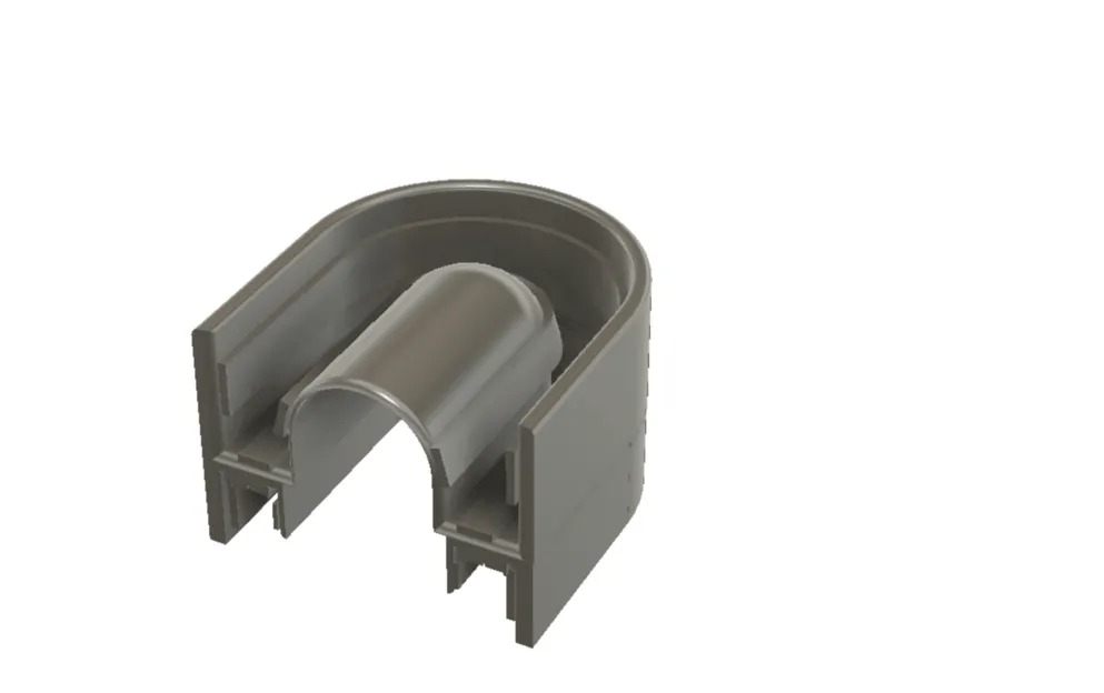


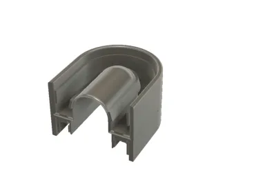
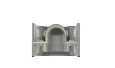
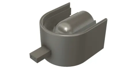
This invention relates to a tray-based three-dimensional oral imaging system designed to capture accurate digital images of a patient’s teeth, gums, and lips in a single, stable scanning process. Unlike conventional handheld scanning wands, the system uses a dental tray integrated with built-in illumination and imaging sensors that remain stationary inside the mouth during scanning, improving consistency and ease of use.
The system addresses common clinical challenges such as patient discomfort, motion-related scanning errors, and moisture buildup on imaging lenses. This is achieved through the integration of airflow or suction channels positioned near the sensors, which help reduce fogging and condensation during use. Optional features include an external attachment for scanning the outer lips and a dual-tray configuration that enables simultaneous scanning of the upper and lower jaws, including the capture of jaw alignment and bite relationship data.
The captured image data is processed into high-quality three-dimensional digital dental models, which can be used for dental diagnosis, treatment planning, orthodontic applications, and the design and manufacture of dental restorations.
Problem Addressed by the Invention
Traditional methods of capturing dental impressions and oral images are often uncomfortable, time-consuming, and prone to errors. Conventional impression materials can cause gag reflex, discomfort, and inaccurate results, while also requiring multiple steps and repeat procedures. Even modern digital solutions have notable limitations.
Most existing digital intraoral scanning systems rely on handheld scanning wands. These devices must be continuously moved inside the mouth, which can cause discomfort for patients and fatigue for clinicians. Because the scanner is always in motion, the quality of the captured images depends heavily on operator skill. Small hand movements, changes in angle, or patient motion can lead to image distortion, stitching errors, or incomplete scans, often requiring rescanning.
Another common issue is moisture inside the mouth. Saliva, fogging, and condensation can easily form on camera lenses, reducing image clarity and making it difficult to capture fine details such as tooth margins or gum lines. Current systems often rely on powders, drying procedures, or repeated cleaning of the scanner tip, increasing procedure time and complexity.
Benchtop scanners are an alternative, but they require physical impressions or models to be made first. These systems are bulky, not chairside-friendly, and add extra steps, delays, and costs to the dental workflow.
Existing systems also struggle to efficiently capture a complete view of the oral region in one step. Scanning the upper teeth, lower teeth, gums, lips, and jaw relationships often requires multiple devices, repositioning, or separate procedures. Capturing accurate jaw alignment and movement data is especially challenging with current tools.
Overall, the dental industry lacks a compact, patient-friendly, and efficient imaging solution that can capture high-quality, full-mouth digital impressions with minimal motion, reduced moisture interference, and consistent accuracy, while also simplifying the workflow for dental professionals.
Addressing a Major Gap in Prior Art
Yes, the invention clearly addresses a major and well-recognized gap in prior oral imaging technologies.
Prior art in digital dental imaging is largely divided into handheld intraoral scanning wands and benchtop scanners, each with inherent limitations. Handheld scanners require continuous manual movement inside the mouth, making scan quality highly dependent on operator skill and patient cooperation. This often leads to motion-related inaccuracies, stitching errors, incomplete scans, and patient discomfort. Benchtop scanners, on the other hand, require physical impressions or models, adding extra steps, time, and cost, and do not support real-time, chairside imaging.
A critical gap in prior art is the lack of a stable, stationary scanning solution that can capture high-quality, full-mouth digital impressions without continuous hand movement. Existing systems also fail to adequately manage moisture, fogging, and condensation near imaging sensors, which directly affects image clarity and margin detection. Many solutions rely on powders, repeated drying, or manual cleaning, none of which are efficient or patient-friendly.
Another significant gap is the inability to capture multiple oral regions simultaneously. Prior art typically requires separate scans for upper teeth, lower teeth, gums, lips, and jaw alignment. Accurate recording of bite position and jaw relationships, especially in a repeatable and clinically reliable manner, remains a challenge with conventional scanning tools.
The invention addresses these gaps by introducing a tray-based oral imaging system where imaging sensors and illumination elements are fixed in position relative to the teeth and gums, eliminating motion-induced errors. The inclusion of integrated air and suction channels directly resolves the moisture and condensation problem at the source. Furthermore, the stacked tray configuration with a jaw-positioning ramp enables simultaneous upper and lower jaw scanning while capturing accurate bite relationships, a capability largely absent in prior systems.
In summary, the invention fills a major gap in prior art by combining stationary full-mouth scanning, active moisture control, and simultaneous jaw relationship capture into a single, integrated system designed for efficient clinical use.
Brief Description of the Gap Addressed
Yes, the invention clearly addresses a major and well-recognized gap in prior oral imaging technologies.
Prior art in digital dental imaging is largely divided into handheld intraoral scanning wands and benchtop scanners, each with inherent limitations. Handheld scanners require continuous manual movement inside the mouth, making scan quality highly dependent on operator skill and patient cooperation. This often leads to motion-related inaccuracies, stitching errors, incomplete scans, and patient discomfort. Benchtop scanners, on the other hand, require physical impressions or models, adding extra steps, time, and cost, and do not support real-time, chairside imaging.
A critical gap in prior art is the lack of a stable, stationary scanning solution that can capture high-quality, full-mouth digital impressions without continuous hand movement. Existing systems also fail to adequately manage moisture, fogging, and condensation near imaging sensors, which directly affects image clarity and margin detection. Many solutions rely on powders, repeated drying, or manual cleaning, none of which are efficient or patient-friendly.
Another significant gap is the inability to capture multiple oral regions simultaneously. Prior art typically requires separate scans for upper teeth, lower teeth, gums, lips, and jaw alignment. Accurate recording of bite position and jaw relationships, especially in a repeatable and clinically reliable manner, remains a challenge with conventional scanning tools.
The invention addresses these gaps by introducing a tray-based oral imaging system where imaging sensors and illumination elements are fixed in position relative to the teeth and gums, eliminating motion-induced errors. The inclusion of integrated air and suction channels directly resolves the moisture and condensation problem at the source. Furthermore, the stacked tray configuration with a jaw-positioning ramp enables simultaneous upper and lower jaw scanning while capturing accurate bite relationships, a capability largely absent in prior systems.
In summary, the invention fills a major gap in prior art by combining stationary full-mouth scanning, active moisture control, and simultaneous jaw relationship capture into a single, integrated system designed for efficient clinical use.
**Unique Features of the Product / Process **
Tray-Based, Stationary Scanning Architecture Uses a U-shaped dental tray with fixed imaging strips, eliminating handheld scanner movement and reducing image distortion and operator dependency.
Integrated Imaging Strips Along Oral Contours Multiple imaging sensors and illumination sources are positioned along inner, outer, and base surfaces of the tray to capture comprehensive oral data simultaneously.
Active Moisture and Fog Control Built-in airflow and suction channels near the sensors actively reduce saliva condensation and fogging without relying on surface powders.
Configurable Sensor Arrangements Imaging sensors can be arranged in condensed or expanded, matched or alternating configurations to adapt to different mouth sizes and imaging needs.
Optional Extraoral Scanning Capability A detachable external extension allows scanning of the lips and outer mouth area in the same imaging session.
Stacked Tray Configuration for Full-Mouth Capture Two trays can be used together to scan upper and lower teeth simultaneously, reducing scanning time and alignment errors.
Jaw Position and Motion Recording A detachable ramp and optional motion sensors enable accurate capture of jaw position and movement during scanning.
Direct Digital Output for CAD/CAM Workflows Captured images are processed into precise 3D digital dental impressions suitable for restorative, orthodontic, and prosthetic applications.
Industries where the invention can be useful?
Industries Where the Invention Can Be Useful – European Countries The tray-based 3D oral imaging system is applicable across a wide range of industries within Europe’s mature and highly regulated dental and healthcare ecosystem. Primary Industries • Dental Clinics & Private Practices General dentistry, orthodontics, implantology, prosthodontics, and cosmetic dentistry clinics using digital impressions. • Hospitals & Public Healthcare Systems Dental, oral surgery, and maxillofacial departments within public and private hospitals. • Dental Clinic Chains / Group Practices Multi-location dental networks operating across EU countries, focused on standardization and efficiency. • Dental Laboratories Labs producing crowns, bridges, dentures, aligners, surgical guides, and implant components using digital workflows. Secondary & Supporting Industries • Dental Equipment Manufacturers Companies developing and commercializing dental imaging, scanning, and digital dentistry hardware. • Dental Equipment Distributors & Resellers Regional and country-level distributors supplying advanced dental technologies across Europe. • Dental Diagnostic & Imaging Centers Specialized centers providing imaging and diagnostic services to clinics and hospitals. • Dental Education & Training Institutions Universities, dental schools, and professional training centers teaching digital dentistry techniques. • Orthodontic Aligner Manufacturers Companies producing clear aligners and orthodontic systems requiring precise 3D oral scans. • CAD/CAM & Dental Software Providers Firms offering design, modeling, and digital workflow software that rely on accurate intraoral scan data. • Medical Device Research & Innovation Centers R&D organizations and innovation hubs working on next-generation dental and medical imaging technologies. Summary The invention is commercially relevant across clinical care, dental manufacturing, education, distribution, and digital dentistry technology development throughout European countries, supporting both private and public healthcare systems.An estimate of the total addressable market?
European digital dentistry market is estimated at ~USD 2.0B–2.6B annually.Potential Customers/End Users. Who might benefit?
Potential Customers / End Users – European Countries Primary End Users • Dental Clinics & Private Practices General dentistry, orthodontic, implant, prosthodontic, and cosmetic clinics using digital impressions and 3D oral imaging. • Hospitals & Public Healthcare Dental Units Dental, oral surgery, and maxillofacial departments within public and private hospitals across Europe. • Dental Clinic Groups & DSOs Multi-location dental chains and group practices seeking standardized, efficient, and operator-independent imaging solutions. Secondary Beneficiaries • Dental Laboratories Labs producing crowns, bridges, dentures, aligners, and surgical guides using digital workflows. • Dental Diagnostic & Imaging Centers Specialized centers providing oral imaging and diagnostic services to clinics and hospitals. • Dental Equipment Manufacturers & Distributors Companies developing, distributing, or integrating dental imaging and digital dentistry hardware across European markets. • Dental Universities & Training Institutions Dental schools, universities, and professional training centers teaching modern digital dentistry workflows. • Orthodontic Aligner & Dental Appliance Manufacturers Companies producing clear aligners and customized dental appliances requiring accurate 3D oral scans. • CAD/CAM & Digital Dentistry Software Providers Software companies relying on precise scan data for dental design and manufacturing. Summary Any organization involved in clinical dental care, dental manufacturing, education, or digital dentistry technology in Europe can directly benefit from this invention’s stable, efficient, and scalable 3D oral imaging approach.Actions
Added all portfolio
| Country | Current Status | Patent Application Number | Patent Number | Applicant / Current Assignee Name | Title | Google Patent Link |
| Europe | Awaiting Examination | MARGHALANI, Thamer | Not mentioned | EP22804127A | THREE-DIMENSIONAL ORAL IMAGING SYSTEM AND METHOD | Not mentioned |
You may also like the following patent
THREE-DIMENSIONAL ORAL IMAGING SYSTEM AND METHOD
| Available For: China | Pharmaceuticals and Biotechnology |
This invention relates to a tray-based three-dimensional oral imaging system designed to capture accurate digital images of a patient’s teeth, gums, and lips in a single, stable scanning...
| Application Number | 2022800333318 |
| Patent Number | |
| Current Status | Awaiting Examination |
THREE-DIMENSIONAL ORAL IMAGING SYSTEM AND METHOD
| Available For: India | Pharmaceuticals and Biotechnology |
This invention relates to a tray-based three-dimensional oral imaging system designed to capture accurate digital images of a patient’s teeth, gums, and lips in a single, stable scanning process....
| Application Number | 202317073670 |
| Patent Number | N/A |
| Current Status | Awaiting Examination |
Smart Medication Gamification System
| Available For: India | Pharmaceuticals and Biotechnology |
A smart medication management system that uses connected pharmaceutical packaging and software to monitor when patients take their medication. The system records medication events, reminds patients...
| Application Number | 201717046644 |
| Patent Number | 579106 |
| Current Status | Granted |
THREE-DIMENSIONAL ORAL IMAGING SYSTEM AND METHOD
| Available For: United States | Pharmaceuticals and Biotechnology |
This invention relates to a three-dimensional oral imaging system that captures accurate digital images of a patient’s teeth, gums, and lips using a specially designed dental tray placed inside the...
| Application Number | 17/324,980 |
| Patent Number | US11382727 |
| Current Status | Granted |
Tarpan glasses
| Available For: India | Pharmaceuticals and Biotechnology |
Tarpan Glasses are a reusable eyewear-based device designed to make the Ayurvedic eye lubrication procedure known as Tarpan easier, faster, and more comfortable. The device creates a sealed space...
| Application Number | 202021004558 |
| Patent Number | 545912 |
| Current Status | Granted |
Motion Artifact reduction in ECG harness
| Available For: India | Pharmaceuticals and Biotechnology |
The patent is for getting a clinical grade ECG during normal movement and exercises (like in a treadmill stress test) conveniently and quickly from a dry electrode wearable ECG harness without...
| Application Number | 85/CHE/2014 |
| Patent Number | 540782 |
| Current Status | Granted |
3D Oral Imaging Tray System
| Available For: Saudi Arabia | Pharmaceuticals and Biotechnology |
This invention relates to a three-dimensional oral imaging system designed to capture accurate digital impressions of teeth, gums, and surrounding oral structures in a simple and comfortable manner....
| Application Number | 523451561 |
| Patent Number | SA 19790 |
| Current Status | Granted |