


THREE-DIMENSIONAL ORAL IMAGING SYSTEM AND METHOD
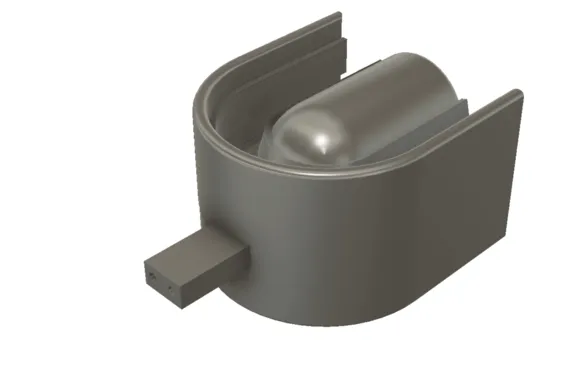
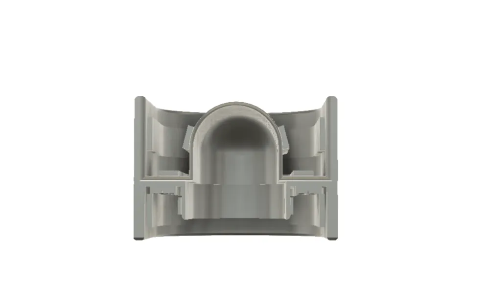
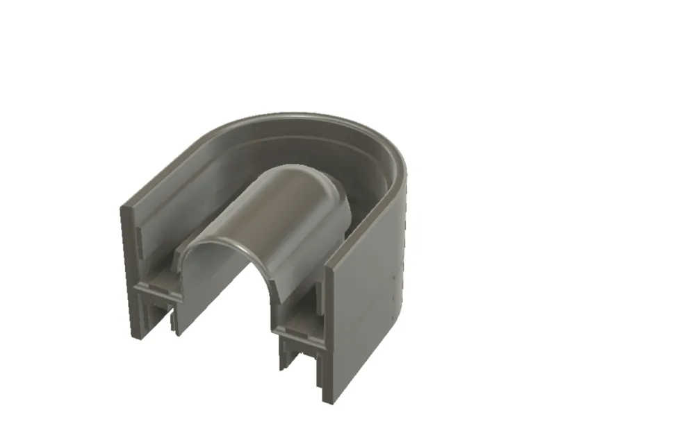


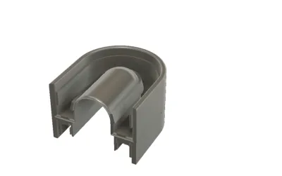
This invention relates to a tray-based three-dimensional oral imaging system designed to capture accurate digital images of a patient’s teeth, gums, and lips in a single, stable scanning process. Unlike conventional handheld scanning wands, the system uses a dental tray integrated with built-in illumination and imaging sensors that remain stationary inside the mouth during scanning, improving consistency and ease of use.
The system addresses common clinical challenges such as patient discomfort, motion-related scanning errors, and moisture buildup on imaging lenses. This is achieved through the integration of airflow or suction channels positioned near the sensors, which help reduce fogging and condensation during use. Optional features include an external attachment for scanning the outer lips and a dual-tray configuration that enables simultaneous scanning of the upper and lower jaws, including the capture of jaw alignment and bite relationship data.
The captured image data is processed into high-quality three-dimensional digital dental models, which can be used for dental diagnosis, treatment planning, orthodontic applications, and the design and manufacture of dental restorations.
Problem Addressed by the Invention
Traditional methods of capturing dental impressions and oral images are often uncomfortable, time-consuming, and prone to errors. Conventional impression materials can cause gag reflex, discomfort, and inaccurate results, while also requiring multiple steps and repeat procedures. Even modern digital solutions have notable limitations.
Most existing digital intraoral scanning systems rely on handheld scanning wands. These devices must be continuously moved inside the mouth, which can cause discomfort for patients and fatigue for clinicians. Because the scanner is always in motion, the quality of the captured images depends heavily on operator skill. Small hand movements, changes in angle, or patient motion can lead to image distortion, stitching errors, or incomplete scans, often requiring rescanning.
Another common issue is moisture inside the mouth. Saliva, fogging, and condensation can easily form on camera lenses, reducing image clarity and making it difficult to capture fine details such as tooth margins or gum lines. Current systems often rely on powders, drying procedures, or repeated cleaning of the scanner tip, increasing procedure time and complexity.
Benchtop scanners are an alternative, but they require physical impressions or models to be made first. These systems are bulky, not chairside-friendly, and add extra steps, delays, and costs to the dental workflow.
Existing systems also struggle to efficiently capture a complete view of the oral region in one step. Scanning the upper teeth, lower teeth, gums, lips, and jaw relationships often requires multiple devices, repositioning, or separate procedures. Capturing accurate jaw alignment and movement data is especially challenging with current tools.
Overall, the dental industry lacks a compact, patient-friendly, and efficient imaging solution that can capture high-quality, full-mouth digital impressions with minimal motion, reduced moisture interference, and consistent accuracy, while also simplifying the workflow for dental professionals.
Addressing a Major Gap in Prior Art
Yes, the invention clearly addresses a major and well-recognized gap in prior oral imaging technologies.
Prior art in digital dental imaging is largely divided into handheld intraoral scanning wands and benchtop scanners, each with inherent limitations. Handheld scanners require continuous manual movement inside the mouth, making scan quality highly dependent on operator skill and patient cooperation. This often leads to motion-related inaccuracies, stitching errors, incomplete scans, and patient discomfort. Benchtop scanners, on the other hand, require physical impressions or models, adding extra steps, time, and cost, and do not support real-time, chairside imaging.
A critical gap in prior art is the lack of a stable, stationary scanning solution that can capture high-quality, full-mouth digital impressions without continuous hand movement. Existing systems also fail to adequately manage moisture, fogging, and condensation near imaging sensors, which directly affects image clarity and margin detection. Many solutions rely on powders, repeated drying, or manual cleaning, none of which are efficient or patient-friendly.
Another significant gap is the inability to capture multiple oral regions simultaneously. Prior art typically requires separate scans for upper teeth, lower teeth, gums, lips, and jaw alignment. Accurate recording of bite position and jaw relationships, especially in a repeatable and clinically reliable manner, remains a challenge with conventional scanning tools.
The invention addresses these gaps by introducing a tray-based oral imaging system where imaging sensors and illumination elements are fixed in position relative to the teeth and gums, eliminating motion-induced errors. The inclusion of integrated air and suction channels directly resolves the moisture and condensation problem at the source. Furthermore, the stacked tray configuration with a jaw-positioning ramp enables simultaneous upper and lower jaw scanning while capturing accurate bite relationships, a capability largely absent in prior systems.
In summary, the invention fills a major gap in prior art by combining stationary full-mouth scanning, active moisture control, and simultaneous jaw relationship capture into a single, integrated system designed for efficient clinical use.
Brief Description of the Gap Addressed
Yes, the invention clearly addresses a major and well-recognized gap in prior oral imaging technologies.
Prior art in digital dental imaging is largely divided into handheld intraoral scanning wands and benchtop scanners, each with inherent limitations. Handheld scanners require continuous manual movement inside the mouth, making scan quality highly dependent on operator skill and patient cooperation. This often leads to motion-related inaccuracies, stitching errors, incomplete scans, and patient discomfort. Benchtop scanners, on the other hand, require physical impressions or models, adding extra steps, time, and cost, and do not support real-time, chairside imaging.
A critical gap in prior art is the lack of a stable, stationary scanning solution that can capture high-quality, full-mouth digital impressions without continuous hand movement. Existing systems also fail to adequately manage moisture, fogging, and condensation near imaging sensors, which directly affects image clarity and margin detection. Many solutions rely on powders, repeated drying, or manual cleaning, none of which are efficient or patient-friendly.
Another significant gap is the inability to capture multiple oral regions simultaneously. Prior art typically requires separate scans for upper teeth, lower teeth, gums, lips, and jaw alignment. Accurate recording of bite position and jaw relationships, especially in a repeatable and clinically reliable manner, remains a challenge with conventional scanning tools.
The invention addresses these gaps by introducing a tray-based oral imaging system where imaging sensors and illumination elements are fixed in position relative to the teeth and gums, eliminating motion-induced errors. The inclusion of integrated air and suction channels directly resolves the moisture and condensation problem at the source. Furthermore, the stacked tray configuration with a jaw-positioning ramp enables simultaneous upper and lower jaw scanning while capturing accurate bite relationships, a capability largely absent in prior systems.
In summary, the invention fills a major gap in prior art by combining stationary full-mouth scanning, active moisture control, and simultaneous jaw relationship capture into a single, integrated system designed for efficient clinical use.
**Unique Features of the Product / Process **
Tray-Based, Stationary Scanning Architecture Uses a U-shaped dental tray with fixed imaging strips, eliminating handheld scanner movement and reducing image distortion and operator dependency.
Integrated Imaging Strips Along Oral Contours Multiple imaging sensors and illumination sources are positioned along inner, outer, and base surfaces of the tray to capture comprehensive oral data simultaneously.
Active Moisture and Fog Control Built-in airflow and suction channels near the sensors actively reduce saliva condensation and fogging without relying on surface powders.
Configurable Sensor Arrangements Imaging sensors can be arranged in condensed or expanded, matched or alternating configurations to adapt to different mouth sizes and imaging needs.
Optional Extraoral Scanning Capability A detachable external extension allows scanning of the lips and outer mouth area in the same imaging session.
Stacked Tray Configuration for Full-Mouth Capture Two trays can be used together to scan upper and lower teeth simultaneously, reducing scanning time and alignment errors.
Jaw Position and Motion Recording A detachable ramp and optional motion sensors enable accurate capture of jaw position and movement during scanning.
Direct Digital Output for CAD/CAM Workflows Captured images are processed into precise 3D digital dental impressions suitable for restorative, orthodontic, and prosthetic applications.
Industries where the invention can be useful?
Commercial and Market Potential (India-Focused) In the Indian dental market, this invention can be positioned as a faster, more consistent, and more comfortable 3D oral scanning alternative to conventional impressions and operator-dependent handheld intraoral scanning—useful for high-volume clinics, aligner workflows, implant planning, and lab-ready digital dentistry. Indian Industries / Segments Where It Can Be Useful • Dental Clinics & Multi-specialty Dental Centers (Tier 1–3 cities) For digital impressions for crowns, bridges, veneers, and general restorative workflows with reduced chair-time. • Dental Clinic Chains / DSOs Standardized scanning across branches (important in India where operator skill varies), enabling consistent quality and faster throughput. • Orthodontics & Clear Aligner Providers (India-based aligner brands + clinics) Full-arch scans for aligner planning and monitoring; faster capture can improve patient conversion and case turnaround. • Implant Centers & Oral Surgery Practices Digital impressions and jaw relation capture support implant prosthetics and full-mouth rehabilitation workflows. • Dental Laboratories (Crown/Bridge Labs, Digital CAD/CAM Labs) Direct digital input for CAD/CAM production and reduced remakes; strong fit for labs serving multiple clinics. • Dental Colleges, Teaching Hospitals & Training Institutions Standardized case documentation, student training, and repeatable scans for academic workflows. • Cosmetic Dentistry & Smile Design Clinics Lip + intraoral imaging capability supports aesthetic planning and patient visualization/communication. • Mobile Dental Clinics / Corporate Dental Camps / Outreach Programs Potential for more predictable scans in “on-site” environments where traditional impressions are slow and messy. • Medical Device Distributors & Dental Equipment Integrators (India Channel Partners) A licensable / marketable product category that can be bundled with CAD/CAM milling and 3D printing ecosystems. • Indian Manufacturing & Private-Label Opportunities Scope to localize components, assembly, and consumables to meet Indian pricing expectations and scale distribution. Why It Fits India Commercially (Non-Technical) • High patient volume + price sensitivity → value comes from speed, fewer retakes, and standardized output. • Uneven operator skill across clinics → tray-based stationary scanning can improve repeatability. • Fast-growing aligner + implant demand → strong pull for reliable 3D digital impression workflows.Potential Customers/End Users. Who might benefit?
1. Core Dental Imaging & Intraoral Scanning Market (India) India has 3+ lakh practicing dentists, with a fast-growing share moving toward digital workflows. Large and mid-sized clinics, dental chains, orthodontic centers, and implant-focused practices are the primary buyers of 3D scanning solutions. The Indian market for digital dental scanners and related imaging devices is estimated to be in the range of USD 250–400 million annually, considering equipment sales, upgrades, and replacements. 2. Downstream Digital Dentistry & CAD/CAM Ecosystem (Influenced Market) Dental labs, aligner manufacturers, implant prosthetic labs, and milling/printing service providers rely on digital impressions. The broader digital dentistry ecosystem in India (CAD/CAM, aligners, implants, prosthetics) is estimated to exceed USD 1–1.5 billion annually, where accurate and standardized scanning is a critical enabling input. This invention directly feeds into and benefits from this expanding ecosystem. 3. Licensing & OEM Opportunity (Device Manufacturers) Indian and international dental device manufacturers, private-label brands, and distributors actively look for cost-effective, differentiating scanning technologies. From a licensing or OEM perspective, the addressable opportunity spans platform licensing, per-unit royalties, and bundled hardware sales, making the effective TAM larger than direct device sales alone. Overall Market Perspective (Simplified) Direct Addressable Market (India): ~USD 250–400 million Influenced / Enablement Market: ~USD 1+ billion Commercial Reality: Even 1–3% penetration represents a meaningful commercial opportunity in the Indian market. Commercial Takeaway (Non-Technical) This invention targets a large, fast-growing Indian dental market where demand is shifting toward faster, standardized, and digital-first solutions. The market size supports product sales, strategic licensing, and manufacturing partnerships, making it attractive for both investors and device companies.Actions
Added all portfolio
| Country | Current Status | Patent Application Number | Patent Number | Applicant / Current Assignee Name | Title | Google Patent Link |
| India | Awaiting Examination | 202317073670 | N/A | MARGHALANI, Thamer | THREE-DIMENSIONAL ORAL IMAGING SYSTEM AND METHOD | Not mentioned |
You may also like the following patent
Motion Artifact reduction in ECG harness
| Available For: India | Pharmaceuticals and Biotechnology |
The patent is for getting a clinical grade ECG during normal movement and exercises (like in a treadmill stress test) conveniently and quickly from a dry electrode wearable ECG harness without...
| Application Number | 85/CHE/2014 |
| Patent Number | 540782 |
| Current Status | Granted |
THREE-DIMENSIONAL ORAL IMAGING SYSTEM AND METHOD
| Available For: United States | Pharmaceuticals and Biotechnology |
This invention relates to a three-dimensional oral imaging system that captures accurate digital images of a patient’s teeth, gums, and lips using a specially designed dental tray placed inside the...
| Application Number | 17/324,980 |
| Patent Number | US11382727 |
| Current Status | Granted |
3D Oral Imaging Tray System
| Available For: Saudi Arabia | Pharmaceuticals and Biotechnology |
This invention relates to a three-dimensional oral imaging system designed to capture accurate digital impressions of teeth, gums, and surrounding oral structures in a simple and comfortable manner....
| Application Number | 523451561 |
| Patent Number | SA 19790 |
| Current Status | Granted |
THREE-DIMENSIONAL ORAL IMAGING SYSTEM AND METHOD
| Available For: Germany | Pharmaceuticals and Biotechnology |
This invention relates to a tray-based three-dimensional oral imaging system designed to capture accurate digital images of a patient’s teeth, gums, and lips in a single, stable scanning...
| Application Number | MARGHALANI, Thamer |
| Patent Number | |
| Current Status | Awaiting Examination |
THREE-DIMENSIONAL ORAL IMAGING SYSTEM AND METHOD
| Available For: China | Pharmaceuticals and Biotechnology |
This invention relates to a tray-based three-dimensional oral imaging system designed to capture accurate digital images of a patient’s teeth, gums, and lips in a single, stable scanning...
| Application Number | 2022800333318 |
| Patent Number | |
| Current Status | Awaiting Examination |
Smart Medication Gamification System
| Available For: India | Pharmaceuticals and Biotechnology |
A smart medication management system that uses connected pharmaceutical packaging and software to monitor when patients take their medication. The system records medication events, reminds patients...
| Application Number | 201717046644 |
| Patent Number | 579106 |
| Current Status | Granted |
Tarpan glasses
| Available For: India | Pharmaceuticals and Biotechnology |
Tarpan Glasses are a reusable eyewear-based device designed to make the Ayurvedic eye lubrication procedure known as Tarpan easier, faster, and more comfortable. The device creates a sealed space...
| Application Number | 202021004558 |
| Patent Number | 545912 |
| Current Status | Granted |