


3D Oral Imaging Tray System


This invention relates to a three-dimensional oral imaging system designed to capture accurate digital impressions of teeth, gums, and surrounding oral structures in a simple and comfortable manner. Instead of using handheld scanners or traditional impression materials, the system uses a dental tray fitted with integrated imaging sensors and light sources that are positioned inside the mouth.
When the tray is placed in the patient’s mouth, the imaging components simultaneously scan multiple areas of the oral cavity, allowing full-mouth or partial-mouth images to be captured in a single procedure. The system also includes features to manage moisture and reduce fogging on the sensors, helping maintain image clarity during scanning. In some configurations, the system can also capture images of the outer lip area at the same time.
The collected imaging data is processed to generate high-quality 3D digital models that can be used for dental diagnosis, treatment planning, orthodontics, implants, and restorative procedures. Overall, the invention provides a more stable, faster, and patient-friendly alternative to conventional dental impression methods and operator-dependent handheld scanners, supporting efficient digital dentistry workflows.
Problem the Invention Addresses (Simplified Explanation)
Modern dentistry relies heavily on accurate dental impressions to plan treatments such as crowns, bridges, orthodontics, implants, and restorative procedures. However, the commonly used methods for capturing these impressions present several practical problems.
Traditional impression techniques use soft, moldable materials placed inside the mouth. These methods are often uncomfortable for patients, can cause gagging, and require significant chair time. They are also sensitive to movement and handling, which can result in distortions or inaccurate impressions. If errors occur, the process must be repeated, increasing time, cost, and patient dissatisfaction.
Digital intraoral scanning was introduced to improve this process, but existing digital solutions also have limitations. Most current systems rely on handheld scanning wands that must be manually moved around the mouth. These scanners are highly operator-dependent and require skill and steady hand movement to capture accurate data. Even small movements by the patient or operator can lead to incomplete scans, stitching errors, or reduced image quality. This makes consistent results difficult, especially in busy clinics or where operator skill levels vary.
Another challenge during intraoral scanning is moisture and fogging. Saliva, humidity, and condensation inside the mouth can collect on camera lenses or sensors, reducing visibility and accuracy. Managing moisture often requires additional tools or repeated interruptions during scanning.
Additionally, many existing systems scan the mouth in segments rather than capturing multiple areas at once. This increases scanning time and can lead to alignment issues between different scanned regions. Capturing both internal oral structures (teeth and gums) and external features (such as lips) usually requires separate steps or equipment.
Overall, the industry lacks a solution that can:
- Capture stable and accurate full-mouth 3D images in a single or reduced number of steps
- Reduce dependency on operator skill and hand movement
- Improve patient comfort during scanning
- Minimize errors caused by moisture and fogging
- Increase efficiency and consistency across different clinics and users
The invention addresses these limitations by rethinking how intraoral imaging is performed, focusing on stability, simultaneous data capture, and improved reliability for everyday dental practice.
The Invention – Solution to the Identified Problem (Simplified Explanation)
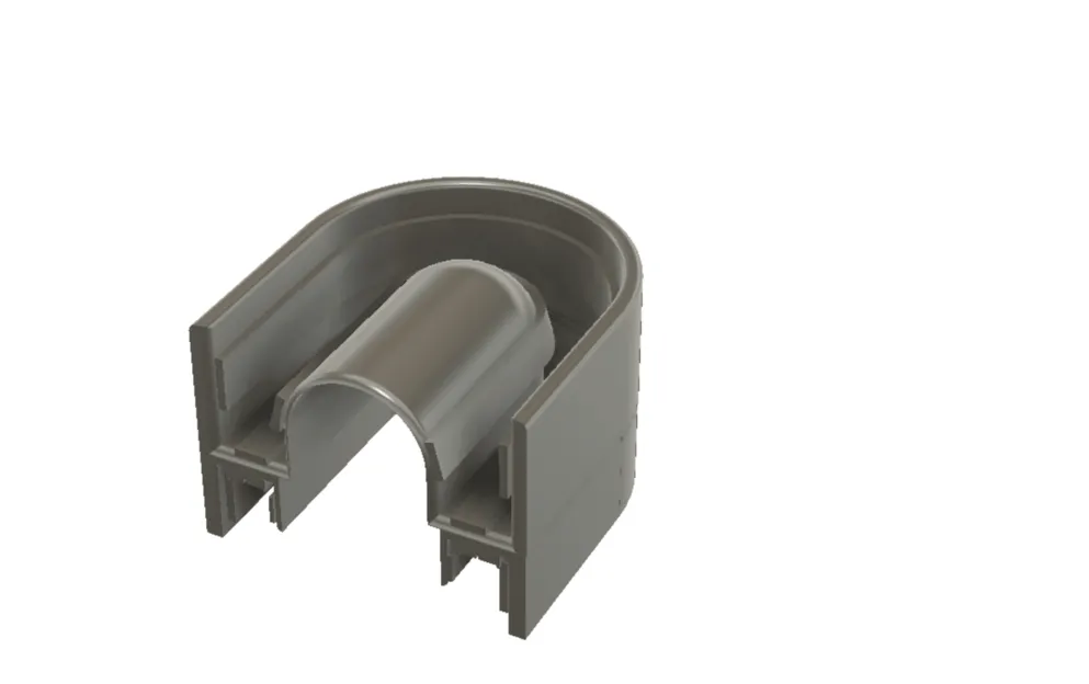
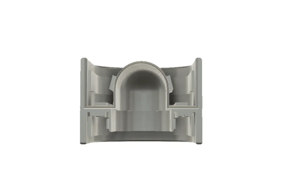
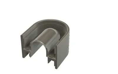
The invention provides a tray-based three-dimensional oral imaging system that offers a more stable, efficient, and patient-friendly way to capture digital dental impressions. Instead of relying on handheld scanners or traditional impression materials, the system uses a dental tray fitted with integrated imaging components that are positioned inside the mouth in a fixed and controlled manner.
When the tray is placed in the patient’s mouth, multiple imaging sensors and light sources embedded along the tray simultaneously capture images of the teeth, gums, and surrounding oral structures. Because the imaging components remain largely stationary relative to the mouth, the system significantly reduces errors caused by hand movement, patient motion, or operator technique. This allows consistent and repeatable image capture across different users and clinical environments.
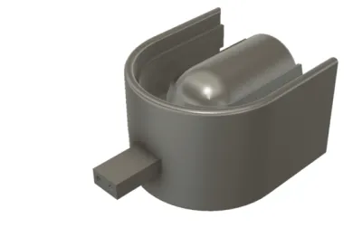
To address common issues related to moisture and fogging, the system incorporates airflow or suction channels positioned near the imaging sensors. These channels help remove saliva, moisture, and condensation from the imaging area, ensuring clearer images without frequent interruptions. This improves reliability during scanning and reduces the need for additional moisture-control tools.
In certain configurations, the system includes external extensions that allow imaging of outer oral features, such as the lips, at the same time as internal scanning. This enables a more complete oral model to be captured in a single procedure rather than through multiple separate steps.
The invention also supports configurations where upper and lower dental trays can be used together, allowing full-mouth scanning in one session. Optional positioning features help align the jaw accurately, supporting reliable bite registration and jaw relationship analysis.
Captured image data is transmitted to a processing system that converts the information into high-quality 3D digital models. These models can be used directly in digital dentistry workflows, including treatment planning, orthodontics, prosthetics, implants, and restorative procedures.
Overall, the invention solves key problems in dental imaging by:
- Reducing dependence on operator skill
- Improving scan stability and consistency
- Enhancing patient comfort
- Minimizing moisture-related image issues
- Enabling faster and more comprehensive data capture
This makes the system well suited for high-volume clinics, standardized dental chains, and modern digital dentistry environments seeking reliable and efficient imaging solutions.
Brief Description of the Gap Addressed
Prior art in dental impression and intraoral imaging generally falls into two main categories:
traditional physical impression methods and digital intraoral scanners, primarily handheld or benchtop systems. Each of these approaches leaves a significant unmet gap that the invention directly addresses.
Traditional impression techniques rely on soft molding materials placed inside the mouth. While widely used, these methods are uncomfortable for patients, time-consuming, prone to distortion, and unsuitable for fully digital workflows. They also require repeat procedures when impressions are inaccurate, increasing clinical inefficiency.
Existing digital solutions improve accuracy but introduce new limitations. Handheld intraoral scanners depend heavily on the operator’s skill and steady hand movement. Small variations in scanning technique, patient motion, or scanning path can lead to stitching errors, incomplete scans, and inconsistent results. This makes it difficult to achieve standardized outcomes across clinics, operators, and patient types. Benchtop scanners, on the other hand, require impressions or models to be created first, adding extra steps and delaying workflows.
Another key gap in prior art is the lack of a stable, fixedurl
fixed-position intraoral imaging solution. Most prior systems capture images sequentially rather than simultaneously, increasing scan time and alignment complexity. In addition, moisture management—such as saliva, fogging, and condensation on imaging sensors—remains a persistent problem in prior systems and is typically handled through external tools rather than being integrated into the imaging device itself.
Furthermore, prior art generally treats internal oral scanning and external facial or lip scanning as separate processes, requiring different devices or multiple procedures. This separation increases time, cost, and complexity for clinics.
The invention addresses this gap by introducing a tray-based, fixed-position, multi-sensor oral imaging system that:
- Captures multiple oral regions simultaneously rather than sequentially
- Reduces operator dependency by stabilizing sensor position inside the mouth
- Integrates moisture and fog control directly into the imaging structure
- Enables internal and external oral imaging in a single coordinated system
- Supports full-arch and full-mouth scanning in fewer steps
By shifting from handheld, motion-dependent scanning to a stable, integrated tray-based imaging architecture, the invention fills a clear and commercially significant gap in prior art, offering improved consistency, efficiency, and scalability for modern digital dentistry workflows.
Unique Features of the Product / Process / Service
• Tray-Based Fixed Imaging Design Uses a dental tray that holds imaging sensors and light sources in a stable, fixed position inside the mouth, reducing reliance on hand movement and operator skill.
• Simultaneous Multi-Area Scanning Captures images of multiple oral regions at the same time, enabling faster and more consistent full-arch or full-mouth scanning compared to sequential scanning methods.
• Integrated Imaging Sensors and Lighting Combines imaging sensors and illumination directly into the tray structure to ensure uniform lighting and reliable image capture across teeth, gums, and surrounding oral structures.
• Built-In Moisture and Fog Control Incorporates airflow or suction channels near the imaging sensors to reduce saliva buildup, fogging, and condensation during scanning, improving image clarity.
• Internal and External Oral Imaging Capability Optional external extensions allow scanning of outer oral features, such as lips, alongside internal dental structures in a coordinated process.
• Upper and Lower Tray Configuration Support Allows the use of upper and lower trays together to capture full-mouth data in a single session, supporting accurate bite and jaw relationship recording.
• Digital Workflow Compatibility Produces high-quality 3D digital models suitable for direct use in modern digital dentistry applications, including diagnosis, treatment planning, orthodontics, and restorative workflows.
These features collectively differentiate the invention from conventional handheld scanners and traditional impression methods by emphasizing stability, integration, and efficiency.
Industries where the invention can be useful?
Commercial and Market Potential This invention has strong commercial potential in Saudi Arabia’s growing dental and healthcare market, driven by increased healthcare investment, adoption of digital dentistry, and demand for high-quality patient care. Key Industries and Use Segments • Dental Clinics & Specialty Practices General, orthodontic, implant, prosthodontic, and cosmetic dental clinics requiring accurate digital impressions. • Hospitals & Medical Centers Dental and maxillofacial departments for diagnosis, treatment planning, and surgical support. • Dental Laboratories Labs producing crowns, bridges, aligners, dentures, and surgical guides using digital workflows. • Dental Clinic Chains / DSOs Multi-location dental networks seeking standardized, efficient, and operator-independent imaging solutions. • Dental Education & Training Institutions Universities and training centers teaching modern digital dentistry techniques. • Dental Equipment Distributors Local distributors and suppliers expanding portfolios with advanced imaging technologies. Market Relevance The system supports faster workflows, consistent results, and improved patient comfort, aligning well with Saudi Arabia’s healthcare modernization and digital dentistry adoption.An estimate of the total addressable market?
Market Size (Total Addressable Market – Saudi Arabia) (Optional estimate) High-level TAM (Saudi dental technology market): • The broader Saudi “digital dentistry” market is estimated at ~USD 1.1B (covers CAD/CAM, imaging, 3D printing, digital workflows, etc.). Relevant TAM for this invention (intraoral 3D scanning / digital impressions): Because this patent is specifically an intraoral 3D imaging / digital impression capture system, a practical TAM proxy is the intraoral scanning + related software/workflow segment within digital dentistry. A reasonable working range for Saudi Arabia is: • ~USD 55M to USD 110M (≈ SAR 206M to SAR 413M) as a subset of the broader digital dentistry spend (assumption: intraoral scanning/digital impression capture is ~5–10% of overall digital dentistry). Sanity-check context (market capacity): • Saudi Arabia’s dental devices market is estimated ~USD 472M in 2025, which indicates significant annual spend on dental equipment and supports the above range as commercially plausible for a focused segment like scanning. • The Ministry of Health reported 3,253 MOH dental clinics (public side alone), indicating a large installed base of potential users in addition to private clinics and chains. (Note: These figures are directional TAM estimates for commercialization planning, not audited market numbers.)Potential Customers/End Users. Who might benefit?
Potential Customers / End Users (Saudi Arabia) Primary End Users • Dental Clinics & Specialist Practices General dentistry, orthodontics, implants, prosthodontics, and cosmetic dentistry clinics that require accurate digital impressions and efficient workflows. • Hospital Dental & Maxillofacial Departments Public and private hospitals using advanced imaging for diagnosis, treatment planning, and surgical procedures. • Dental Clinic Chains / DSOs Multi-location dental groups seeking standardized, operator-independent imaging across branches. Secondary Beneficiaries • Dental Laboratories Labs producing crowns, bridges, aligners, dentures, and surgical guides that rely on high-quality 3D digital models. • Dental Diagnostic & Imaging Centers Centers offering specialized imaging services to multiple clinics. • Dental Education & Training Institutions Universities and training centers teaching digital dentistry and modern clinical workflows. • Dental Equipment Distributors & Integrators Companies supplying advanced dental technologies to clinics and hospitals across Saudi Arabia. Summary Any organization involved in diagnosis, treatment planning, restorative dentistry, orthodontics, or digital dental manufacturing can directly benefit from this invention’s stable, efficient, and patient-friendly 3D oral imaging approach.Actions
Added all portfolio
| Country | Current Status | Patent Application Number | Patent Number | Applicant / Current Assignee Name | Title | Google Patent Link |
| Saudi Arabia | Granted | 523451561 | SA 19790 | MARGHALANI, Thamer | THREE-DIMENSIONAL ORAL IMAGING SYSTEM AND METHOD | Not mentioned |
You may also like the following patent
THREE-DIMENSIONAL ORAL IMAGING SYSTEM AND METHOD
| Available For: India | Pharmaceuticals and Biotechnology |
This invention relates to a tray-based three-dimensional oral imaging system designed to capture accurate digital images of a patient’s teeth, gums, and lips in a single, stable scanning process....
| Application Number | 202317073670 |
| Patent Number | N/A |
| Current Status | Awaiting Examination |
Tarpan glasses
| Available For: India | Pharmaceuticals and Biotechnology |
Tarpan Glasses are a reusable eyewear-based device designed to make the Ayurvedic eye lubrication procedure known as Tarpan easier, faster, and more comfortable. The device creates a sealed space...
| Application Number | 202021004558 |
| Patent Number | 545912 |
| Current Status | Granted |
Smart Medication Gamification System
| Available For: India | Pharmaceuticals and Biotechnology |
A smart medication management system that uses connected pharmaceutical packaging and software to monitor when patients take their medication. The system records medication events, reminds patients...
| Application Number | 201717046644 |
| Patent Number | 579106 |
| Current Status | Granted |
THREE-DIMENSIONAL ORAL IMAGING SYSTEM AND METHOD
| Available For: United States | Pharmaceuticals and Biotechnology |
This invention relates to a three-dimensional oral imaging system that captures accurate digital images of a patient’s teeth, gums, and lips using a specially designed dental tray placed inside the...
| Application Number | 17/324,980 |
| Patent Number | US11382727 |
| Current Status | Granted |
Motion Artifact reduction in ECG harness
| Available For: India | Pharmaceuticals and Biotechnology |
The patent is for getting a clinical grade ECG during normal movement and exercises (like in a treadmill stress test) conveniently and quickly from a dry electrode wearable ECG harness without...
| Application Number | 85/CHE/2014 |
| Patent Number | 540782 |
| Current Status | Granted |
THREE-DIMENSIONAL ORAL IMAGING SYSTEM AND METHOD
| Available For: China | Pharmaceuticals and Biotechnology |
This invention relates to a tray-based three-dimensional oral imaging system designed to capture accurate digital images of a patient’s teeth, gums, and lips in a single, stable scanning...
| Application Number | 2022800333318 |
| Patent Number | |
| Current Status | Awaiting Examination |
THREE-DIMENSIONAL ORAL IMAGING SYSTEM AND METHOD
| Available For: Germany | Pharmaceuticals and Biotechnology |
This invention relates to a tray-based three-dimensional oral imaging system designed to capture accurate digital images of a patient’s teeth, gums, and lips in a single, stable scanning...
| Application Number | MARGHALANI, Thamer |
| Patent Number | |
| Current Status | Awaiting Examination |